CIB provides anatomical models that help you visualise and understand the human body in three dimensions. These professional, highly detailed models allow you to view anatomy from a completely new perspective. They are not only a scientific resource, but also offer the opportunity to learn through observation, analysis and hands-on work with realistic representations of the human body.
These models will help students understand the relationships between organs, muscles, bones and the nervous system, and will support topographic anatomy learning during preparation for tests, exams and practical sessions. Learning in 3D helps students retain information more quickly and develop their anatomical imagination, which is crucial in medical education.
Check out the models available and make use of them whilst studying on your own in the library.
Guidelines for the use of anatomical models PDF
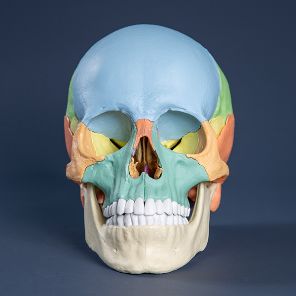
Osteopathic skull A realistic skull model that can be dismantled into 22 parts, with clearly marked bones and joints. It allows for a detailed study of the structure of individual components. It helps in learning the anatomy of the skull bones, identifying their parts and understanding their spatial relationships.
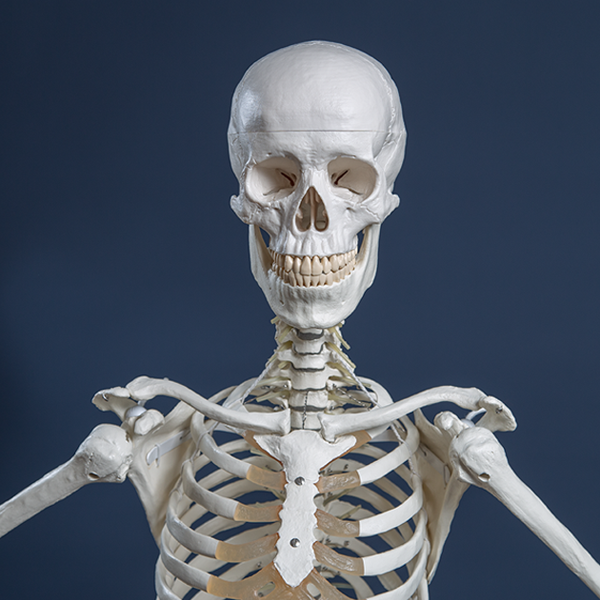
Skeleton “Hugo” with movable spine A full-size skeleton model with a movable spine and joints, and detachable limbs. It facilitates the study of the skeletal system and the mechanics of movement – particularly useful for understanding how joints work and the biomechanics of the body.
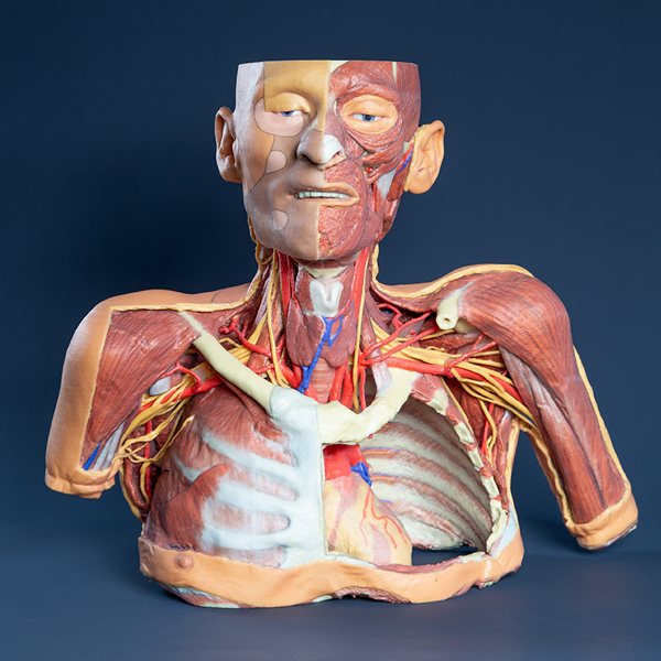
Head, Neck and Shoulder with angiosomes A detailed model depicting muscles, blood vessels, nerve supply and organs in a realistic arrangement. It helps in learning regional anatomy and understanding the relationships between the nervous, circulatory and muscular systems.
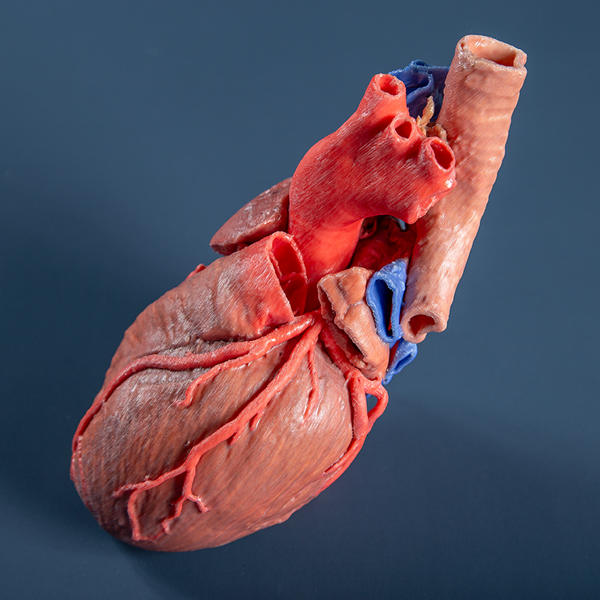
Heart and the distal trachea, carina and primary bronchi A precise representation of the heart, including the major blood vessels and parts of the trachea and bronchi. It helps students learn about the anatomy of the heart, blood circulation and the topography of structures within the mediastinum.
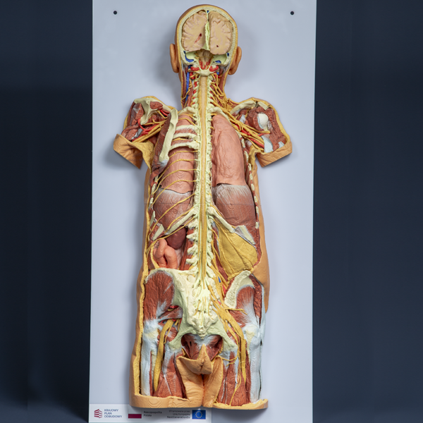
Nervous System Dissection (posterior view) A model showing the central and peripheral nervous systems in a dorsal cross-section, with the spinal cord and nerves clearly visible. It helps to understand the course of the nerves and their relationship to other anatomical structures.
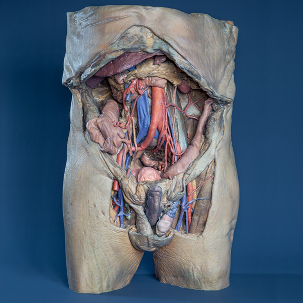
Abdomen with bilateral Hernias A torso model illustrating the abdominal organs and various types of inguinal hernias. It helps in learning the anatomy of the internal organs and understanding the pathology and mechanism of hernia formation.